Khalifa University researchers find a way to use convolutional neural networks to identify cancer in tissue samples, which could speed up diagnosis and improve outcomes in patients with colorectal cancer.
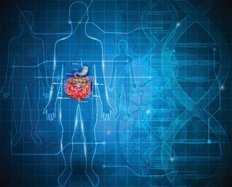
Colorectal cancer is the third most common cancer among men and women worldwide, and the second most common cause of cancer-related mortality. Most colorectal cancers are due to old age and lifestyle factors, with only a small number of cases due to underlying genetic disorders. It typically starts as a benign tumor, such as a polyp, which over time becomes cancerous. Like all forms of cancer, early diagnosis and differentiation of the tumor are crucial for a patient’s survival and wellbeing.
Colorectal cancer may be diagnosed by obtaining a sample of the colon and using histopathology – the study of changes in tissues caused by diseases – to determine the characteristics of the tumour tissue at the microscopic level.
Histology is the study of the microanatomy of cells, tissues and organs as seen through a microscope. The structure of each tissue in the body is directly related to its function and diseases affect tissues in distinctive ways. Studying the histology of a tissue can be very useful in making a diagnosis and determining the severity and progress of a condition.
Because of the great variety of tests that are available, and the high level of skill needed to carry out and interpret them, researchers are beginning to turn to computational pathology and artificial intelligence techniques to identify in tissue samples diseases like cancer.
Dr. Sajid Javed, Assistant Professor, and Dr. Naoufel Werghi, Associate Professor of Electrical Engineering and Computer Science, have collaborated with researchers from around the world to develop algorithms to identify samples of colorectal cancer tissue. A paper based on this research has been published in Medical Image Analysis.
“Computational pathology is a fast-growing research area in cancer diagnosis and can play an instrumental role in helping medical professionals detect and classify tumors,” said Dr. Javed.
Cancer histology reveals underlying molecular processes and disease progression and contains rich phenotypic information that is predictive of patient outcomes.
The phenotype is the set of observable characteristics or traits of an organism or a tissue. Image-based phenotyping aims to develop the computer vision techniques and tools needed to recover quantitative data from a wide range of images. But phenotyping presents challenging problems, particularly in images from colorectal cancer tissues.
Aided by advances in slide scanning microscopes and computing, convolutional neural networks (CNNs) have emerged as an important image analysis tool. CNNs use a network of interconnected layers of filters that highlight important patterns in the images and can continue to learn from previous results.
“Manual examination of tissue samples is time-consuming, highly subjective, and often affected by the observer,” explained Dr. Javed. “Meanwhile, algorithms analyzing digitized Whole Slide Images (WSIs) can examine hundreds of thousands of cells and billions of pixels to differentiate seven distinct tissue phenotypes.”
Deep learning methods require large amounts of annotated histology data for training, which may be tedious to obtain. Additionally, while these methods may be effective in determining tumor tissue, the tissues in colorectal cancer also contain a rich mix of several other types of tissue, including smooth muscle, inflammatory, necrotic, and benign tissue. Any algorithm must be taught to distinguish between these tissue types to be effective.
Texture analysis is a commonly used approach for tissue phenotyping, where texture features are computed to train classifiers, which are then used to predict distinct tissue types.
“Texture analysis may be attractive due to its simplicity but it does not fully capture the biological diversity of tissue components,” explained Dr. Javed.
“Recent methods have proposed integrating cellular connectivity features, which are used as a proxy to cellular interaction features. The notion of cellular connectivity features is based on the fact that spatially adjacent cells have a higher probability of receiving inter-cellular signals from each other than from cells that are farther away. It has also been shown that inter-cellular signals between various types of cells can influence the progression of cancer. However, a dynamic network of tumor growth cannot be adequately modelled by a single type of interaction. Our technique uses a multiplex network model to represent the intricate relationships between cell populations. We propose four different types of cellular networks integrating a variety of features representing tissue characteristics at different levels.”
In the researchers’ model, cells from a WSI are detected and classified into five distinct categories using a deep neural network. Then, four different types of cellular interaction features are computed and used to construct a four-layer multiplex graph. Since each slide contains thousands of cells, the slides are segmented into tiles or patches, which helps the algorithm determine the distribution of different types of tissues across the cells.
“There are many directions in which this work can be further extended,” added Dr. Javed. “Further cellular types such as blood cells could improve performance and also reveal more micro-level tissue communities. Additionally, our framework could be adapted to WSIs of different types of cancer. Of course, in clinical practice, our work can help medical practitioners understand the contents of the WSI and make more accurate and timely diagnoses.”
This work was funded by the Khalifa University of Science and Technology and the UK Medical Research Council. The collaborators were also supported by the PathLAKE digital pathology consortium, which is funded from the Data to Early Diagnosis and Precision Medicine strand of the UK government’s Industrial Strategy Challenge Fund, managed and delivered by UK Research and Innovation.
Jade Sterling
Science Writer
5 July 2021