The Medical Imaging Laboratory at Khalifa University has recently acquired the world’s latest spectral photon-counting computed tomography (CT) scanner (MARS Microlab 5X120). This equipment is the first of its type to be used in the GCC region to support laboratory/pre-clinical research.
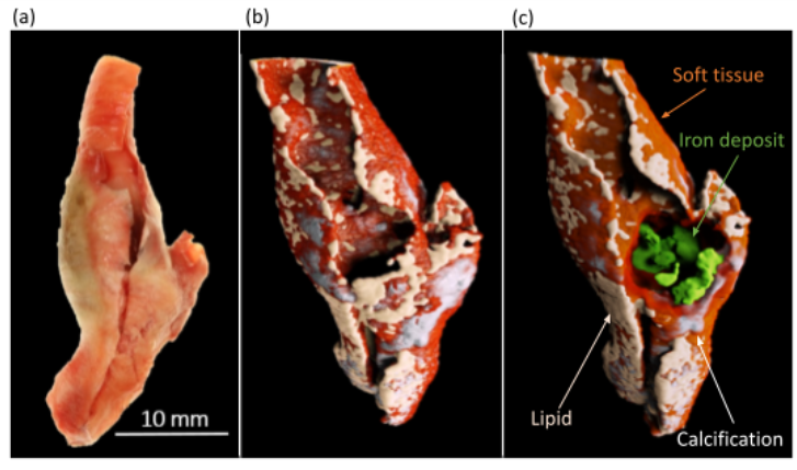
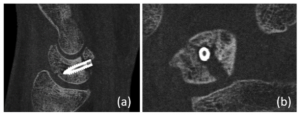
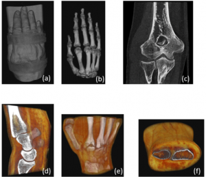
The micro-CT research scanner uses x-ray energies relevant to the human diagnostic imaging range (20–140 keV) and a photon-processing detector that processes the energy of individually detected x-ray photons. The scanner provides high-resolution (<90-micron voxel size) non-destructive three-dimensional multi-energy CT images that are decomposed into material images based on the density and atomic composition of each material. The scanner can accommodate in vitro, in vivo, and excised biological samples sizes of up to 125 mm in diameter and 450 mm in length.

The scanner is intended to be used for medical physics teaching and biomedical imaging research purposes. The Medical Imaging Laboratory is envisaged to become an imaging hub, providing a platform for local and international researchers that want to gain a better understanding of the use of scanner technology for their research. Technological advantages of the medical imaging platform in general and Mars photon-counting CT, in particular, will coincide with Khalifa University’s goal to contribute toward creating a knowledge-based sustainable economy in Abu Dhabi. The applications of the technology include, but are not limited to, developing artificial intelligence-based material reconstruction and metal artefacts reduction software, characterization of biosecurity and geological samples, development of dental and metal implants, exploring the development of targeted high-atomic number nanoparticles with theranostic capabilities, drug delivery quantification and characterization of biological specimens such as bone health, arthritis, cancer imaging and cardiovascular disease.

For more information and collaborative interest, please contact Dr. Aamir Raja (aamir.raja@ku.ac.ae).
Erica Solomon
Senior Publication Specialist
25 May 2022