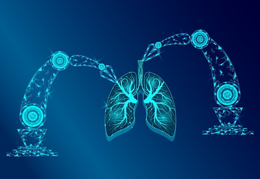
Khalifa University’s Dr. Peter Corridon has advanced tissue engineering with the development of bioengineered scaffolds made from ‘decellularized’ mouse, rat, pig, camel and sheep tissue segments, such as blood vessels, trachea, esophagi, and whole organs like the kidney and eye that may be used as replacement tissues and organs . His research is among the first to evaluate the integrity of bioartificial blood vessels and whole organs under human physiological conditions, examining how they function over time and how they can be extended to make any decellularized architecture less susceptible to degradation and more viable for long-term transplants.
Read the Arabic story here: https://researchku.com/news-extended/234
Taking organs from animals and stripping the cells from the blood vessels could be the new solution to treating medical problems, including retinopathy, amputations, and kidney failure.
After this cleaning process, all that remains is a web of collagen and protein called the extracellular matrix, which gives the blood vessel its structure. This is tissue engineering, and it forms the basis of research from Khalifa University focused on designing scaffolds for tissue and organ regrowth in patients with diseases that lead to organ failure.
Dr. Peter Corridon, Assistant Professor of Physiology and Immunology at Khalifa University, investigated the integrity of vascular networks in decellularized tissues to support the development of blood vessels for kidneys. The results of this study, published in Nature Scientific Reports, wil aid in implementing lifesaving treatments for conditions including diabetes-induced kidney failure. Indeed, the first person to receive a bioengineered blood vessel implant was a patient with late-stage kidney disease in 2013. Earlier this month, a US man became the first person in the world to get a heart transplant from a genetically-modified pig.
Diabetes is the leading cause of kidney disease, with about one-third of diabetic adults suffering. The kidneys function to filter wastes and water out of the blood, helping to control blood pressure and maintain a healthy balance of water, salts and minerals in the blood. Blood flows into the kidney through the renal artery, is filtered in the functional units of the kidney, called nephrons, by clusters of tiny blood vessels called glomeruli, and then flows out of the kidney through the renal vein. This occurs throughout the day, with kidneys filtering around 150 quarts of blood every day.
Over time, poorly controlled diabetes can cause damage to the blood vessels in the kidneys, eyes, legs, and feet leading to uncontrolled damage and high blood pressure. High blood pressure can cause further organ damage by increasing the pressure in the delicate capillary systems. Severe damage to these blood vessel clusters can lead to diabetic nephropathy, retinopathy and amputations.
“By the end of this year, it is expected that 30 percent of the adult population in the United Arab Emirates will be diabetic,” Dr. Corridon said. “Almost half of those with diabetes develop significant vascular complications, which can lead to chronic conditions and even end-stage organ failure. These are substantial public health problems, highlighting the need for safe, effective, and innovative ways to treat the underlying conditions of vascular dysfunction.”
For the kidney specifically, traditional methods of treating renal problems include dialysis and transplantation; while dialysis can replace lost filtration capacities, a kidney transplant is the only way to restore all kidney function. However, there is a severe global shortage of transplantable kidneys and other organs. This, coupled with the issue of organ rejection, accentuate the demand for alternative solutions.
“Recent findings suggest that one possible way of addressing this growing issue is to develop replacement blood vessels, which could be used to treat those needing surgical intervention within the UAE,” Dr. Corridon said.
Bioengineered scaffolds can be used to develop bioartificial blood vessels known as human acellular vessels. They are a scaffold for the body to incorporate and provide a platform for cell growth, tunable to each recipient. They also act immediately as blood vessels, allowing the flow of blood through the kidneys while the body’s own cells grow into the matrix.
However, there are circumstances that limit scaffold viability. Dr. Corridon investigated a simplified model to analyze conditions needed to prepare more durable scaffolds for long-term transplantation.
He is developing his scaffolds using decellularized large and small animals to achieve an accurate biomimetic vascular architecture and functionality.
Decellularization is the process of taking an existing natural organ, either from a human or a nonhuman animal donor, and sterilizing it to the extent that only the collage network base remains, forming a natural scaffold. The decellularized scaffold can then be repopulated with a patient’s own cells to produce a personalized tissue.
These porcine scaffolds were subjected to a continuous blood flow at normal human physiological rates through the arteries to examine any dynamic changes in flow through the vessels and to determine their structure.
“Few studies have evaluated the integrity and function of the decellularized vasculature in whole porcine kidneys under physiological conditions,” Dr. Corridon explained. “The majority of these studies have primarily focused on demonstrating the preservation of structure and patency after decellularization and implantation.”
Under normal conditions, the kidneys autoregulate blood flow to maintain blood pressure through the delicate smaller vessels in the glomeruli. Decellularized kidneys, and kidneys in vitro, however, are incapable of autoregulation – meaning, they would be damaged under higher flow rates.
In this study, rates of 500ml/minute and 650ml/minute were used to represent the amount of blood each kidney would receive during resting conditions. The decellularized kidneys suffered damage at these levels, presumably due to their inability to autoregulate, which suggests that the elastin and collagen fibers in the scaffold would be damaged. In comparison, native kidneys possessed ‘sufficient structural barriers’ that prevented comparable damage, even though they were affected by the continuous flow of unfiltered and unreplenished blood.
“What’s important is that the perfusion process, which is the process of bathing an organ or tissue with a fluid, damaged the internal structures of both native and decellularized organs,” Dr. Corridon said. “While a significant difference was observed between perfused and non-perfused native kidneys, no significant difference was detected between perfused native and decellularized organs when perfused at the same rate.”
These findings reveal that the decellularized organs Dr. Corridon developed behave similarly to the native organs in disease conditions.
Dr. Corridon’s study provides a means to investigate how these blood vessels function over time and can be extended to other platforms to identify ways to make any decellularized architecture less susceptible to degradation and more viable for long-term transplantation.
Decellularization technologies hold great promise for the bioartificial tissue and organ industry, and understanding the limitations of these scaffolds will provide insight into the biomechanical improvements needed to increase their quality and support their clinical utility.
Jade Sterling
Science Writer
21 January 2022