New research shows that long-term speech problems following a stroke are caused by co-occurring damage to white matter, not by damage in Broca’s area alone
Read Arabic story here.
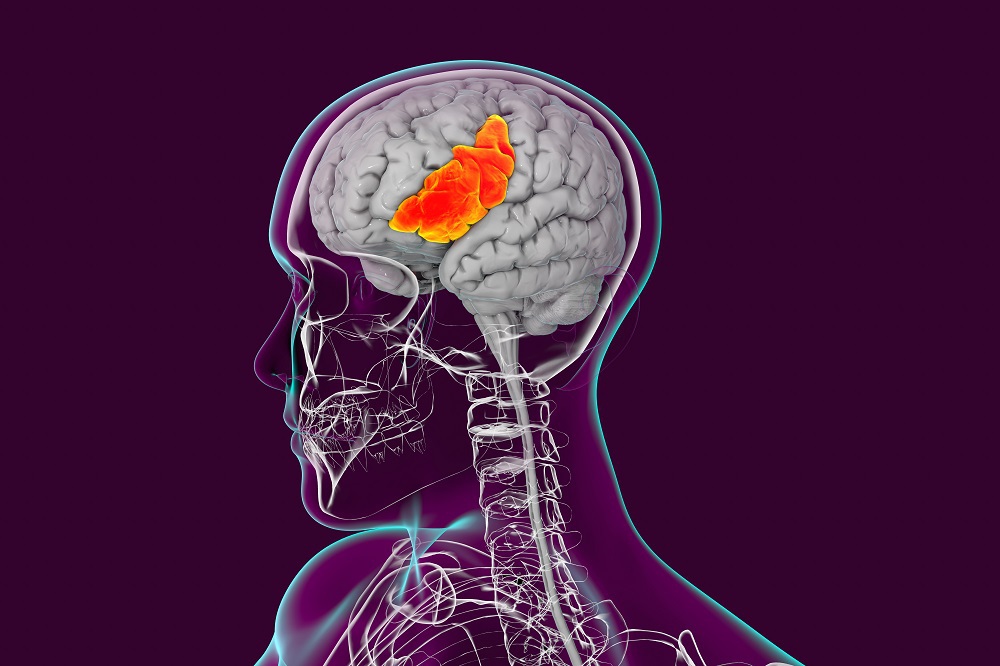
There are several areas of the brain understood to play a critical role in speech and language. One of these is Broca’s area, which is located in the left hemisphere and is associated with speech production and articulation.
The human ability to articulate ideas, as well as use words accurately in spoken and written language, has been attributed to Broca’s area, which is named after the French physician who discovered it in 1861 – Pierre Paul Broca. His work on the left frontal lobe of the brain revealed that the brains of people suffering from aphasia – a loss of language affecting a person’s ability to speak, read and write, which is brought about by neurological damage caused by a stroke – contained legions in a particular part of the cortex, now named Broca’s area. This was the first anatomical proof of localization of brain function.
Given the importance of Broca’s area for language processing abilities, it was widely accepted that damage to this area impairs speech production.
However, an international team that included researchers from University College London (UK), Universidad del Desarrollo (Chile), and Dr. Mohamed Seghier, Professor in KU’s Department of Biomedical Engineering, have challenged the long-held assumption that damage to Broca’s area contributes to long-term speech production impairments after a stroke.
Through their investigation, they discovered that long-term aphasia is not caused by damage to Broca’s area but to damage to neighboring regions, due to the large functional connectivity of this area with adjacent frontal and subcortical areas. They published their work in the journal Brain.
“For over 150 years, clinical aphasiology and behavioral neurology have been influenced by Broca’s finding that stroke survivors with severe and persistent speech impairments had damage to Broca’s area,” explained Dr. Seghier.
“Broca was not able to define the exact extent of the lesions in his patients because, being aware of their historical relevance, he decided not to dissect the specimens but preserve them for future research. His descriptions, therefore, focused on the parts of the lesions that were visible to him, without evaluating the potential contribution of neighboring damage, for example, to the underlying white matter and surrounding cortical areas. It was not until 2007 that the full extent of the lesions incurred by Broca’s two famous cases was revealed in an MRI study showing damage to multiple subcortical grey and white matter regions.”
The research team’s findings confirm that the degree of damage in the surrounding brain tissue – the white and grey matter regions – rather than damage to Broca’s area, is associated with long term aphasia.
White matter is the tissue through which messages pass between different areas of the central nervous system, including the brain. Grey matter contains most of the brain’s neuronal cell bodies and is involved in muscle control and sensory perception such as seeing and hearing, speech and decision making.
The research team investigated their hypothesis by examining whether speech production impediments were worse in stroke survivors who had damage to Broca’s area but not surrounding regions, or who had damage to both Broca’s area and surrounding regions.
Prior research has found that the overwhelming majority of Broca’s aphasia patients present extended brain damage, significantly exceeding the Broca’s area, leading researchers to conclude that lesions restricted to Broca’s area are associated with just mild language production defects. A stroke will typically damage multiple neighboring brain regions to the Broca’s area, and in all cases of stroke it is difficult to determine which part of the lesion site is driving the observed behavioral deficits.
To tackle this problem, the research team studied a large number of stroke survivors who all had left frontal lobe damage but differed in the degree of damage to Broca’s area and surrounding areas. Their selection of brain areas was based on a combination of anatomical and functional evidence and the white matter linking the different areas of the brain responsible for speech.
“Unlike previous studies, our analyses were aimed at disentangling how speech production abilities, months after a stroke on the left frontal lobe, were affected by damage to Broca’s area and the degree to which such effects were influenced by damage also occurring to a specific set of neighboring regions,” explained Dr. Seghier. “The team wanted to test whether damage to Broca’s area contributes to speech production impairments that persist for at least three months after a left frontal lobe stroke.”
The team’s work found that evidence in favor of damage to Broca’s area not explaining variance in speech production abilities was eight times stronger than the alternative. The researchers concluded this is positive evidence for the absence of a unique long-lasting effect of Broca’s area damage on speech production abilities. The absence of evidence became evidence of absence, showing that the prior association between Broca’s area damage and long-lasting speech impairments can be attributed to co-occurring damage to white matter.
Jade Sterling
Science Writer
29 April 2021