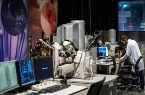
Imaging facility
A host of imaging techniques are available at CMAT that can be utilized for membrane characterization as well as process analysis. Visualization of the membrane surface and cross-section at the macro-, micro- and nano- scale can provide essential information regarding the fabrication process and the surface modification carried out. These tools can be used to determine the presence of materials within the membrane polymer matrix, as well as demonstrate their interaction with the polymer in shaping the membrane characteristics such as pore size and tortuosity.
Imaging tools can also provide a great platform for analyzing membrane fouling. This can be either destructive analysis, such as with fluorescence microscopy for analyzing biofilms through tracking of bacteria and extra-cellular polymeric substances in the film through dyes. In-situ, non-invasive analysis is also possible through tools such as the optical coherence tomography (OCT), which can provide a real- time insight into the development of fouling patterns on the membrane surface.
Some of the noteworthy equipment under this facility are:
- Tecnai TEM 200kV and Titan TEM 300kV (FEI, USA): The Titan enables sub-Angstrom, atomic scale discovery and exploration in both TEM and STEM modes over a wide range of materials and operating conditions.
- Nova Nano SEM 30 Series (FEI, USA): Samples, including the most non-conducting or contaminating materials, can equally be characterized or analyzed in the Nova NanoSEM series, using its unique low vacuum capabilities.
- Quanta 3D FIB SEM (FEI, USA): This is a “dual beam” scanning electron microscope (SEM) that is also equipped with a focused ion beam (FIB). This SEM functions permit microscopic observations of a specimen while the FIB functions allow cutting the sample.
- Optical Coherence Tomography (OCT) Imaging System (Thorlabs GmbH, Germany): Real time mentoring of fouling development on membrane surfaces.